We use cookies to understand how you use our site and to improve your experience. This includes personalizing content and advertising. To learn more, click here. By continuing to use our site, you accept our use of cookies. Cookie Policy.

Features Partner Sites Information LinkXpress hp
Sign In
Advertise with Us
GLOBETECH PUBLISHING LLC

GE Healthcare

GE Healthcare provides medical imaging and information technologies, medical diagnostics, patient monitoring systems,... read more Featured Products: More products

Download Mobile App


Model-Based Iterative Reconstruction Tool Designed to Transform Imaging Technologies

By MedImaging International staff writers
Posted on 19 Feb 2013
Print article
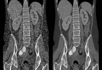
Image: The image at left demonstrates filtered back projection (FBP) image reconstruction using conventional CT imaging, and, at right, an image reconstruction using Veo. Developed by Purdue University, University of Notre Dame and GE Healthcare, Veo is a new CT scanning technology that enables physicians to diagnose patients with high clarity images at previously unattainable low radiation dose levels (Photo courtesy of Purdue University).
Scientists are improving the performance of technologies spanning from medical computed tomography (CT) scanners to digital cameras by utilizing a series of models to mine specific information from very large data pools and then reconstructing the images similar to a jigsaw puzzle.

The new application is called model-based iterative reconstruction (MBIR). “It’s more-or-less how humans solve problems by trial and error, assessing probability and discarding extraneous information,” said Dr. Charles Bouman, Purdue University’s (West Lafayette, IN, USA) professor of electrical and computer engineering and a professor of biomedical engineering.

MBIR has been used in new CT scanning technology that only exposes patients to one-fourth the radiation of conventional CT scanners. In consumer electronics, a new camera has been designed that allows the user to focus the picture after it has been taken. “These innovations are the result of 20 years of research globally to develop iterative reconstruction,” Dr. Bouman said. “We are just scratching the surface. As the research community builds more accurate models, we can extract more information to get better results.”

Radiation exposure reduction in medical CT scanners is due to increased efficiency achieved using the algorithms and models. MBIR decreased “noise” in the data, providing greater clarity that allows the radiologist or radiologic technician to scan the patient at a lower dosage, Dr. Bouman noted. “It’s like having night-vision goggles,” he said. “They enable you to see in very low light, just as MBIR allows you to take high-quality CT scans with a low-power X-ray source.”

Researchers also have applied the application to improve the quality of images captured with an electron microscope. New findings are detailed in a research paper being presented during the Electronic Imaging 2013 Conference held in February 2013, in San Francisco (CA, USA).
Conventionally, imaging sensors and software are designed to detect and measure a specific property. The new approach does the inverse, collecting huge quantities of data and later culling specific information from this pool of information using specialized models and algorithms. “We abandon the idea of purity—collecting precisely what we need,” Dr. Bouman said. “Instead, let’s take all the measurements we possibly can and then later extract what we want. This increases the envelope of what you can do enormously.”

Purdue, the University of Notre Dame (West Bend, IN, USA), and GE Healthcare (Chalfont St. Giles, UK) employed MBIR to create Veo, a new CT scanning technology that enables physicians to diagnose patients with high-clarity images at previously unattainable low radiation dose levels. The technology has been shown to reduce radiation exposure by 78%. “If you can get diagnostically usable scans at such low dosages this opens up the potential to do large-scale screening for things like lung cancer,” Dr. Bouman said. “You open up entirely new clinical applications because the dosage is so low.”

A CT scanner is much better at diagnosing disease than planar X-rays because it provides a three-dimensional (3D) image of the tissue. However, conventional CT scanners emit too much radiation to merit wider diagnostic use. “But as the dosage goes down, the risk-benefit tradeoff for screening will become much more favorable,” Dr. Bouman said. “For electron microscopy, the principle advantage is higher resolutions, but there is also some advantage in reduction of electron dosage, which can damage the sample.”

The research to develop Veo has been a team effort with Dr. Ken Sauer, an associate professor of electrical engineering at Notre Dame, in collaboration with Dr. Jean-Baptiste Thibault, Dr. Jiang Hsieh, and Dr. Zhou Yu. Drs. Thibault and Yu worked on the technology as graduate assistants under Dr. Bouman and Dr. Sauer and both currently work for GE Healthcare. “And, there are lots of other people doing similar and related research at other universities and research labs around the world,” Dr. Bouman said. “Ultimately, 3D X-ray CT images might require little more dosage than old-fashion planar chest X-rays. This would allow CT to be used for medical screening without significant adverse effects.”

Improved resolution could help researchers design the next generation of nanocomposites for applications such as fuel cells and transparent coatings. An article on the technology was published in January 2013 in the journal Current Radiological Reports.

The models and algorithms in MBIR apply probability computations to extract the accurate data, much as people use logical assumptions to draw conclusions. “You search all possible data to find what you are looking for,” Dr. Bouman said. “This is how people solve problems. You saw Bob yesterday at the store; you wonder where he was coming from. Well, you determine that he was probably coming from work because you have some probabilistic models in your mind. You know he probably wasn’t coming from San Francisco because Bob doesn’t go to San Francisco very often, etc.”

MBIR also could bring more cost-effective CT scanners for airport screening. In traditional scanners, an X-ray source rotates at high speeds circling around a chamber, capturing cross-sectional images of luggage positioned inside the chamber. However, MBIR could enable the devices to be streamlined by eliminating the need for the rotating mechanism.

Related Links:

Purdue University
University of Notre Dame
GE Healthcare


New
Stereotactic QA Phantom
StereoPHAN
MRI System
Ingenia Prodiva 1.5T CS
New
Radiation Shielding
Oversize Thyroid Shield
Radiology Software
DxWorks

Print article

Channels

Ultrasound

view channel
Image: Ultrasound detection of vascular changes post-RT corresponds to shifts in the immune microenvironment (Photo courtesy of Theranostics, DOI:10.7150/thno.97759)

Ultrasound Imaging Non-Invasively Tracks Tumor Response to Radiation and Immunotherapy

While immunotherapy holds promise in the fight against triple-negative breast cancer, many patients fail to respond to current treatments. A major challenge has been predicting and monitoring how individual... Read more

Nuclear Medicine

view channel
Image: [18F]3F4AP in a human subject after mild incomplete spinal cord injury (Photo courtesy of The Journal of Nuclear Medicine, DOI:10.2967/jnumed.124.268242)

Novel PET Technique Visualizes Spinal Cord Injuries to Predict Recovery

Each year, around 18,000 individuals in the United States experience spinal cord injuries, leading to severe mobility loss that often results in a lifelong battle to regain independence and improve quality of life.... Read more

General/Advanced Imaging

view channel
Image: The rugged and miniaturized CT scanner is being designed for use beyond a typical hospital setting (Photo courtesy of Micro-X)

World’s First Mobile Whole-Body CT Scanner to Provide Diagnostics at POC

Conventional CT scanners dominate the global medical imaging market, holding approximately 30% of the market share. These scanners are the current standard for various diagnostic applications, including... Read more

Imaging IT

view channel
Image: The new Medical Imaging Suite makes healthcare imaging data more accessible, interoperable and useful (Photo courtesy of Google Cloud)

New Google Cloud Medical Imaging Suite Makes Imaging Healthcare Data More Accessible

Medical imaging is a critical tool used to diagnose patients, and there are billions of medical images scanned globally each year. Imaging data accounts for about 90% of all healthcare data1 and, until... Read more
Copyright © 2000-2025 Globetech Media. All rights reserved.