Expo
view channel
view channel
view channel
view channel
view channel
view channel
view channel
RadiographyMRIUltrasoundNuclear Medicine
Imaging ITIndustry News
Events

- Injury Prediction Rule Reduces Radiographic Imaging Exposure in Children
- AI Detects More Breast Cancers with Fewer False Positives
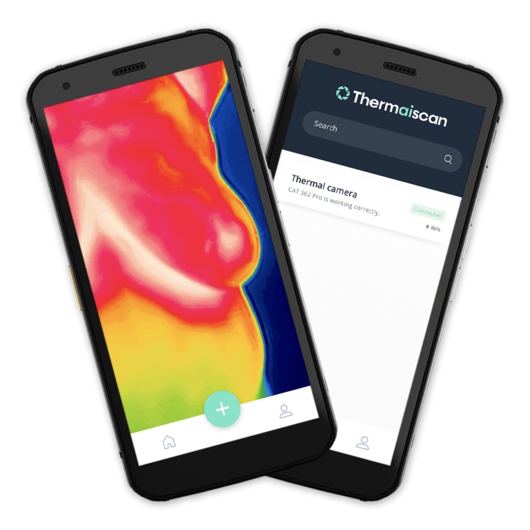
- AI-Powered Portable Thermal Imaging Solution Could Complement Mammography for Breast Cancer Screening
- Novel Breast Imaging System Proves As Effective As Mammography
- AI Assistance Improves Breast-Cancer Screening by Reducing False Positives
- Biology-Driven Radiomics Approach to Identify Rectal Cancer Patients without Tumor Post Therapy
- New Eye Tracking Controlled VR System Enhances MRI Scans for Young Children
- AI Outperforms Radiologists in Detecting Prostate Cancer on MRI
- Non-Invasive Medical Imaging Test Predicts Dementia Nine Years before Diagnosis
- Self-Powered Sensor to Make MRIs More Efficient
- New Radiotracer Generates High Quality and Readily Interpretable Images of Cardiac Amyloidosis
- New PET Radiotracer Enables Same-Day Imaging of Key Gastrointestinal Cancer Biomarker
- New PET Biomarker Predicts Success of Immune Checkpoint Blockade Therapy
- New PET Agent Rapidly and Accurately Visualizes Lesions in Clear Cell Renal Cell Carcinoma Patients
- New Imaging Technique Monitors Inflammation Disorders without Radiation Exposure
- Ultrasound Technology Breaks Blood-Brain Barrier for Glioblastoma Treatment
- Implantable Ultrasound Device Could Replace Electrodes for Deep Brain Stimulation
- Robotic Ultrasound Systems to Assist Doctors during Surgery
- Functional Ultrasound Imaging Records Brain Activity through Transparent Skull Implant
- Ultrasound Wireless Charging To Power Deep Implantable Biomedical Devices
- Artificial Intelligence Tool Enhances Usability of Medical Images
- New AI Tool Accurately Detects Six Different Cancer Types on Whole-Body PET/CT Scans
- Innovative Imaging Technique Helps Assess Bone Loss after Bariatric Surgery
- Imaging Software Improves Lung Diagnosis in Patients Allergic To Medical Contrast Dye
- Bone Density Test Uses Existing CT Images to Predict Fractures
- Global AI in Medical Diagnostics Market to Be Driven by Demand for Image Recognition in Radiology
- AI-Based Mammography Triage Software Helps Dramatically Improve Interpretation Process
- Artificial Intelligence (AI) Program Accurately Predicts Lung Cancer Risk from CT Images
- Image Management Platform Streamlines Treatment Plans
- AI Technology for Detecting Breast Cancer Receives CE Mark Approval
- Hologic Acquires UK-Based Breast Surgical Guidance Company Endomagnetics Ltd.
- Bayer and Google Partner on New AI Product for Radiologists
- Samsung and Bracco Enter Into New Diagnostic Ultrasound Technology Agreement
- IBA Acquires Radcal to Expand Medical Imaging Quality Assurance Offering
- International Societies Suggest Key Considerations for AI Radiology Tools

Expo
 view channel
view channel
view channel
view channel
view channel
view channel
RadiographyMRIUltrasoundNuclear Medicine
Imaging ITIndustry News
Events
Advertise with Us
view channel
view channel
view channel
view channel
view channel
view channel
view channel
RadiographyMRIUltrasoundNuclear Medicine
Imaging ITIndustry News
Events
Advertise with Us


- Injury Prediction Rule Reduces Radiographic Imaging Exposure in Children
- AI Detects More Breast Cancers with Fewer False Positives
- AI-Powered Portable Thermal Imaging Solution Could Complement Mammography for Breast Cancer Screening
- Novel Breast Imaging System Proves As Effective As Mammography
- AI Assistance Improves Breast-Cancer Screening by Reducing False Positives
- Biology-Driven Radiomics Approach to Identify Rectal Cancer Patients without Tumor Post Therapy
- New Eye Tracking Controlled VR System Enhances MRI Scans for Young Children
- AI Outperforms Radiologists in Detecting Prostate Cancer on MRI
- Non-Invasive Medical Imaging Test Predicts Dementia Nine Years before Diagnosis
- Self-Powered Sensor to Make MRIs More Efficient
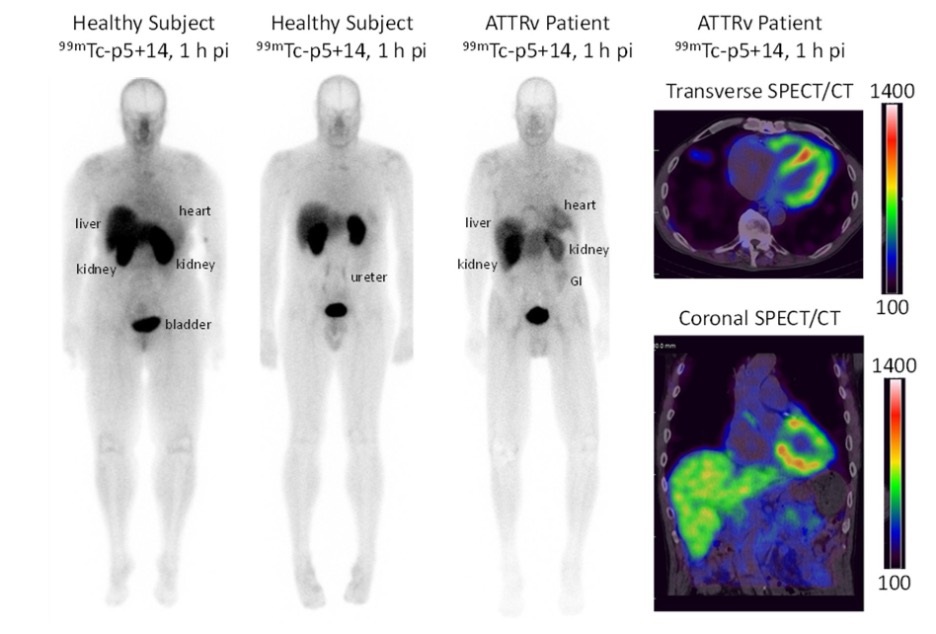
- New Radiotracer Generates High Quality and Readily Interpretable Images of Cardiac Amyloidosis
- New PET Radiotracer Enables Same-Day Imaging of Key Gastrointestinal Cancer Biomarker
- New PET Biomarker Predicts Success of Immune Checkpoint Blockade Therapy
- New PET Agent Rapidly and Accurately Visualizes Lesions in Clear Cell Renal Cell Carcinoma Patients
- New Imaging Technique Monitors Inflammation Disorders without Radiation Exposure
- Ultrasound Technology Breaks Blood-Brain Barrier for Glioblastoma Treatment
- Implantable Ultrasound Device Could Replace Electrodes for Deep Brain Stimulation
- Robotic Ultrasound Systems to Assist Doctors during Surgery
- Functional Ultrasound Imaging Records Brain Activity through Transparent Skull Implant
- Ultrasound Wireless Charging To Power Deep Implantable Biomedical Devices
- Artificial Intelligence Tool Enhances Usability of Medical Images
- New AI Tool Accurately Detects Six Different Cancer Types on Whole-Body PET/CT Scans
- Innovative Imaging Technique Helps Assess Bone Loss after Bariatric Surgery
- Imaging Software Improves Lung Diagnosis in Patients Allergic To Medical Contrast Dye
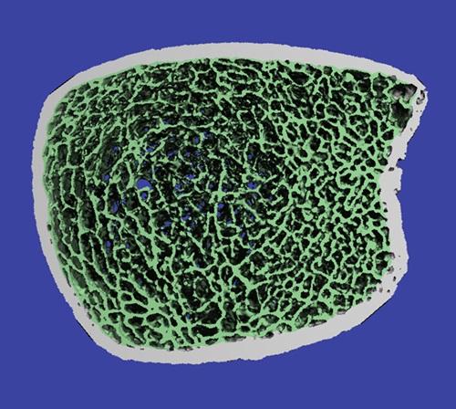
- Bone Density Test Uses Existing CT Images to Predict Fractures
- Global AI in Medical Diagnostics Market to Be Driven by Demand for Image Recognition in Radiology
- AI-Based Mammography Triage Software Helps Dramatically Improve Interpretation Process
- Artificial Intelligence (AI) Program Accurately Predicts Lung Cancer Risk from CT Images
- Image Management Platform Streamlines Treatment Plans
- AI Technology for Detecting Breast Cancer Receives CE Mark Approval
- Hologic Acquires UK-Based Breast Surgical Guidance Company Endomagnetics Ltd.
- Bayer and Google Partner on New AI Product for Radiologists
- Samsung and Bracco Enter Into New Diagnostic Ultrasound Technology Agreement
- IBA Acquires Radcal to Expand Medical Imaging Quality Assurance Offering
- International Societies Suggest Key Considerations for AI Radiology Tools








.jpg)
